Timing
Sudden changes raise different concerns than slow changes. Minutes to hours is a different risk profile than months. Fluctuating symptoms can point toward surface and focusing drivers, but they can also coexist with structural disease.
Definitions, typical symptom patterns, what clinicians evaluate, and red flags that warrant urgent care.

Eye symptoms overlap. The goal here is a practical encyclopedia: what common conditions mean, what symptom patterns are typical, and what information changes clinical decision-making.
This page is organized by category rather than by single symptom, because the same symptom can have multiple causes. For example, fluctuating blur may be driven by tear film instability, focusing strain, or lens changes. A red eye may be allergy, a surface irritation, or a deeper inflammatory problem. The differentiators are pattern, timing, associated symptoms, and exam findings.
For readers trying to navigate real-world care, the most useful mindset is this: clinicians are rarely deciding between hundreds of diseases. They are narrowing possibilities based on a short set of high-signal questions and a focused exam. The sections below explain those decision points.
Symptoms that should not be treated as routine dryness or lifestyle issues.
Many eye complaints are comfort-driven and fluctuate with screens, sleep, and environment. Some presentations are different. They can represent time-sensitive disease where earlier treatment changes outcomes. When uncertain, urgent evaluation is often the safer choice.
If you are trying to decide who to call, what to bring, or how to describe the timeline, see the Care Guide. It is designed for the practical logistics that influence how quickly care happens.
The short list of questions and tests that do most of the sorting.
Most eye evaluations start with a few high-signal dimensions: is the issue painful or not, is vision affected or not, is it one eye or both, and is the change sudden or gradual. Those dimensions shape urgency and narrow the likely categories before advanced testing is even considered.
Sudden changes raise different concerns than slow changes. Minutes to hours is a different risk profile than months. Fluctuating symptoms can point toward surface and focusing drivers, but they can also coexist with structural disease.
Discomfort is common with dryness and allergies. True pain with marked light sensitivity can suggest corneal inflammation, deeper inflammation (uveitis), or pressure emergencies. Context matters, especially contact lens wear.
Clinicians distinguish blur that improves with blinking or rest from blur that does not. They also distinguish central distortion (macula) from peripheral field loss (optic nerve and retina).
For a plain-language explanation of one of the most important modern tests, see OCT explained: the scan that changed eye care.
A high-frequency cause of irritation, fluctuating blur, and visual fatigue.
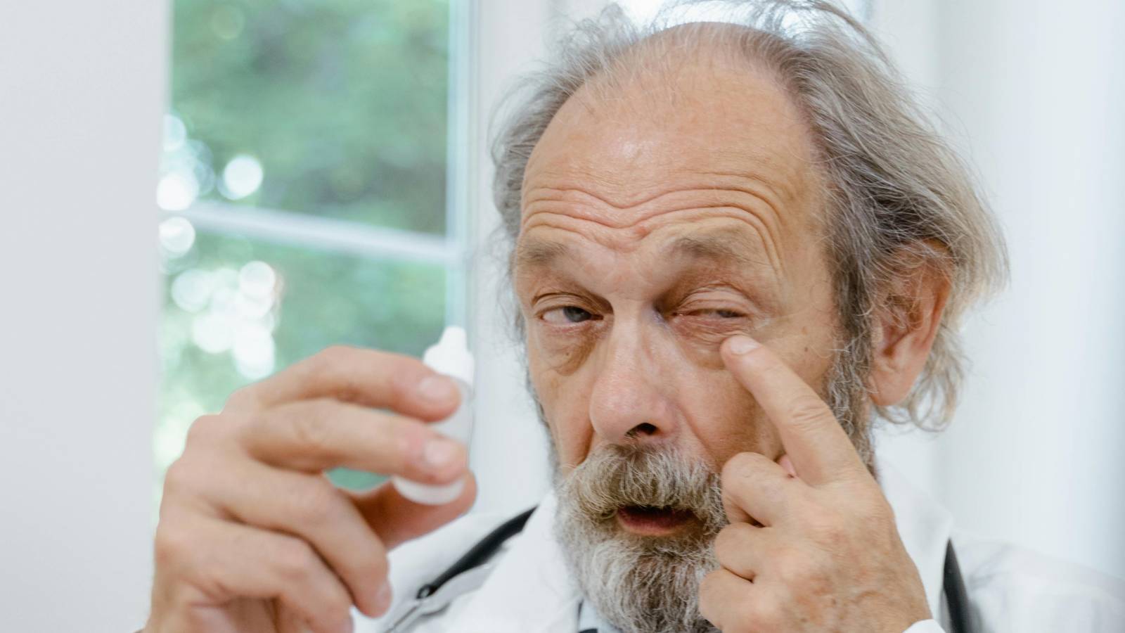
Dry eye is often described as "not enough tears," but that framing is incomplete. A more useful description is tear film instability. The tear film is the first optical surface of the eye. When it breaks up quickly, clarity can fluctuate and the surface can become inflamed. Symptoms often feel inconsistent because the system degrades under load and partially resets with blinking and rest.
Common drivers include reduced blink rate and incomplete blinking during near work, meibomian gland dysfunction (oil layer problems that increase evaporation), environmental dryness and airflow, and medication or systemic contributors. Dry eye is often a systems problem, which is why a single product rarely solves every case.

Lid margin health, meibomian gland function, ocular surface staining, tear breakup time, and contributing factors like blepharitis, medication effects, autoimmune disease, and environmental exposures.
Pain, significant light sensitivity, marked redness, reduced vision, or symptoms that do not match typical dryness patterns warrant evaluation. In contact lens wearers, corneal infection risk changes the threshold.
For deeper routine logic, see Dry eye routines: what they target and why they help. For workstation and screen drivers, see Digital Life.
Glasses, focusing demand, binocular alignment, and why symptoms can appear even when vision seems "fine."
Refractive error describes how the eye focuses light: nearsightedness, farsightedness, and astigmatism. Small refractive errors can be tolerated until the system is stressed by long near work, fatigue, or poor lighting. Symptoms can include headaches, eye fatigue, intermittent blur, and difficulty switching between near and far.
A related category is accommodative and binocular strain. Even with sharp focus, the eyes may work harder to align and converge during near tasks. Symptoms can include soreness around the eyes, intermittent double vision, or a sense of "effort" to keep things single and clear.
For a careful myth vs mechanism breakdown of screens, see Blue light myths: what matters and what does not.
Glare, reduced contrast, and gradual changes that glasses do not fully fix.
The natural lens changes with age. Early lens aging often shows up as reduced focusing ability (presbyopia), followed by progressive optical degradation that can include glare, reduced contrast, dimmer vision, and color changes. When lens clouding and optical scatter become clinically significant, it is called a cataract.
Cataract progression is usually gradual. Many people notice night driving glare first, or a sense that lighting is "not as good as it used to be." Glasses can improve refractive blur, but they cannot remove lens scatter. This is why cataract symptoms are often described as quality-of-vision problems, not just sharpness problems.

The practical question is often functional: is daily life affected enough to justify surgery now. Visual acuity is part of the picture, but glare and contrast complaints can be equally important to real-world function.
See Procedures for a neutral overview of how surgery is done and how lens options are discussed.
Often silent until late, monitored using structure and function tests.
Glaucoma is a group of diseases characterized by progressive optic nerve damage and corresponding visual field loss. Intraocular pressure is an important risk factor, but pressure alone does not define glaucoma. Some people with elevated pressure never develop glaucomatous damage, and some people develop glaucoma with pressures that are not dramatically high.
A key challenge is that central vision can remain good until significant peripheral field has been lost. This is why screening and monitoring matter. Management is often about slowing progression and preserving function over decades rather than fixing a symptom that is obvious early.
Optic nerve exam and OCT imaging of retinal nerve fiber layers. These help detect and trend structural change over time.
Visual field testing measures functional sensitivity. Trend over time is often more useful than a single test result.
A painful red eye with nausea and halos can be an emergency and is not the same as typical chronic glaucoma progression.
Imaging is a major part of modern monitoring. If OCT is unfamiliar, see OCT explained: the scan that changed eye care.
Often influenced by systemic health, frequently monitored with imaging, sometimes treated on schedules.
The retina converts light into neural signals. Because it is highly vascular and metabolically active, it is vulnerable to systemic disease. This category includes age-related macular degeneration (AMD), diabetic retinopathy, retinal vascular occlusions, and time-sensitive emergencies like retinal detachment.
A macular disease affecting central retina. Early stages can be subtle. Later stages can affect central detail vision. Risk discussions often include smoking, age, and genetics. Certain supplement formulas are stage-specific.
Vascular damage associated with diabetes over time. It can progress without symptoms early, which is why regular exams matter. In practice, glucose control and blood pressure influence long-term risk.
New flashes, a sudden increase in floaters, or a curtain-like shadow can be warning signs. Early evaluation can change outcomes.
Retina treatments are often time-structured and data-driven. For a plain-language explanation of common therapy, see Retina injections in plain language.
For systemic risk context, see Nutrition. For care logistics and second opinions, see Second opinions: a practical playbook.
Red eyes are common. Risk depends on pain, vision, light sensitivity, and contact lens history.
"Red eye" is a symptom, not a diagnosis. Conjunctivitis (viral or allergic) is common and often self-limited, but certain presentations are higher risk. Pain, light sensitivity, reduced vision, or contact lens wear changes the risk profile because corneal disease can progress quickly.
Deeper inflammation inside the eye, such as uveitis, can cause pain with light sensitivity, blurred vision, and sometimes floaters. It is not the same category as routine seasonal allergy. Management depends on cause and severity, and evaluation focuses on where inflammation is located and what might be driving it.
If you want a narrative-style example of why second opinions and deeper testing sometimes matter, see When the first visit misses it: Stephanie’s story.
Short definitions for terms commonly used in eye care discussions.
Optical coherence tomography. A non-contact imaging scan that shows cross-sections of retina or optic nerve layers. Used for monitoring and trend tracking.
Intraocular pressure. A risk factor for glaucoma and other conditions, interpreted in context with nerve appearance and testing.
A test of functional sensitivity across vision, often used to monitor glaucoma and neurologic patterns.
Central retina responsible for sharp detail vision. Distortion or central blur raises different questions than peripheral symptoms.
Inflammation inside the eye. Often associated with pain and light sensitivity and can require prompt treatment and workup.
The optical focusing state of the eye. Includes nearsightedness, farsightedness, and astigmatism. Corrected by glasses, contacts, or surgery.
Reputable starting points for definitions, patient education, and evidence discovery.
Patient education on common eye conditions, prevention, and anatomy.
Eye health explanations plus clinical terminology context.
Biomedical literature search maintained by the U.S. National Library of Medicine.